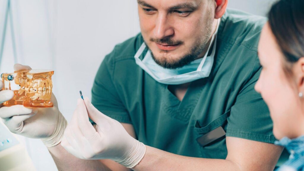
Zygomatic implants exist because the upper jaw does not always give a surgeon enough bone to work with. When the maxilla has resorbed too far for conventional implants, the zygomatic bone (the cheekbone) can serve as a stable anchor point instead. That single design shift, anchoring in the cheekbone rather than the jaw ridge, is what makes full-arch restoration possible for patients who would otherwise be turned away.
Zygomatic implants suit patients with severe upper jaw bone loss whose facial anatomy provides a sound zygomatic bone for anchorage, but the approach is not appropriate for everyone, and candidacy depends on detailed imaging, health screening, and surgical judgment. Facial structure varies enough from person to person that two patients with similar bone loss can receive very different treatment recommendations.
This matters because zygomatic implant surgery works close to the sinus and the orbital floor. For patients exploring zygomatic dental implants in Chula Vista, CA, getting candidacy right, before any incision is made, is the difference between a resolved case and a complicated one.
How Zygomatic Implants Use Facial Anatomy
Zygomatic implants work by shifting anchorage away from the resorbed maxilla and into the zygomatic bone, a dense structure that tooth loss does not affect. The implants are longer than conventional dental implants and follow a trajectory that passes through or alongside the maxillary sinus before engaging the cheekbone itself.
Why the Cheekbone Can Support Implants When the Upper Jaw Cannot
The zygomatic bone stays structurally intact even after decades of tooth loss. Unlike the alveolar ridge, which resorbs once teeth are gone, the cheekbone is not a tooth-bearing structure, so it retains its density and volume regardless of how much maxillary bone has disappeared.
That density gives the implant a solid anchor point. A standard implant placed into severely atrophied maxillary bone has little to grip; a zygomatic implant bypasses that problem by reaching into bone that was never going to resorb.
How the Maxilla, Sinuses, and Cheekbones Affect Implant Placement
Placement depends on the relationship between the maxillary sinus wall, the remaining alveolar ridge, and the zygomatic buttress. Surgeons classify this relationship because it determines whether an implant can travel fully outside the sinus, partly through it, or through its full width.
The Zygoma Anatomy-Guided Approach (ZAGA) formalized this into a classification system based on the maxillary sinus wall’s shape and position. It treats each patient’s anatomy as the starting point for implant trajectory, rather than applying one fixed surgical pathway to every case. Anatomy-based systems such as the Z-Classification extend this idea, factoring in ridge, sinus, and zygoma relationships together to guide surgical decisions.
Who May Qualify for This Advanced Approach
Zygomatic implants are generally considered for patients with advanced maxillary bone loss, classified at the severe end of resorption scales, where conventional implants and even bone grafting are unlikely to succeed. Qualification also depends on sinus anatomy, prior surgical history, and overall health, confirmed through imaging and screening before any surgical plan is finalized.
Which Bone-Loss Patterns May Call for Zygomatic Support
Cawood class V and VI maxillary defects, where the ridge has flattened or resorbed into basal bone, are the pattern most associated with zygomatic candidacy. At this stage, there is often not enough vertical or horizontal bone left to place standard implants without extensive grafting.
Patients who have worn removable dentures for many years frequently fall into this category, since long-term denture use accelerates ridge resorption.
When Previous Implant Failure, Trauma, or Disease Changes the Plan
A history of failed conventional implants, maxillofacial trauma, or disease affecting the maxilla can shift a patient toward the zygomatic approach even without extreme bone loss. Tumor resection, congenital defects, and radiation therapy to the jaw can all leave bone that will not reliably support standard fixtures.
In these cases, the zygomatic bone offers a stable alternative anchor point that sidesteps compromised or missing maxillary structure.
Why Some Patients Are Not Suitable Candidates
Not every patient with severe bone loss qualifies. Active sinus disease, certain anatomical variations in the zygomatic bone itself, uncontrolled systemic conditions, and insufficient zygomatic bone volume can all rule out the procedure.
Smoking, poorly controlled diabetes, and a history of radiation to the surgical field are factors that surgeons weigh carefully, since they affect healing and long-term implant stability.
How CBCT Scans and Health Screening Confirm Eligibility
Cone-beam computed tomography (CBCT) is the imaging standard for confirming zygomatic implant candidacy. It gives the surgical team a three-dimensional view of bone volume, sinus anatomy, and the zygomatic bone’s shape, which two-dimensional X-rays cannot provide.
Health screening runs alongside imaging: medical history, sinus health, and medication use are reviewed before a surgeon commits to a zygomatic plan over a conventional one.
Treatment Trade-Offs, Risks, and Specialist Planning
Zygomatic implants trade a more complex surgery for the ability to skip bone grafting and shorten overall treatment time. That trade-off makes sense for patients with severe atrophy, but it comes with anatomy-specific risks that require an experienced surgical team to manage.
How Zygomatic Implants Compare With Bone Grafting and Conventional Implants
Bone grafting can rebuild a resorbed maxilla enough for conventional implants, but it adds months of healing and carries its own failure risk. Zygomatic implants skip that step by anchoring directly into existing cheekbone.
|
Factor |
Zygomatic Implants |
Bone Grafting + Conventional Implants |
|
Candidate profile |
Severe maxillary atrophy |
Moderate bone loss |
|
Treatment timeline |
Weeks to a few months |
Often 6-12+ months |
|
Additional grafting needed |
Rarely |
Yes, by design |
|
Surgical complexity |
High |
Moderate |
|
Proximity to sinus/orbit |
Close |
Minimal |
What Surgical Risks Are Shaped by Facial Anatomy
Sinusitis is the most commonly reported complication, since the implant path runs close to or through the maxillary sinus. Mucositis and localized gingival recession have also been documented in follow-up studies, though reported rates of severe complications stay lower than other options for extreme upper jaw atrophy.
Anatomical proximity to the orbital floor and infraorbital nerve raises the stakes of imprecise placement, which is why preoperative mapping of these structures is treated as non-negotiable.
Why Experience With Complex Full-Arch Cases Matters
Surgeons who regularly manage complex full-arch and atrophic maxilla cases develop judgment that generic implant experience does not provide. The ITI Consensus Report on zygomatic implants points to surgical technique and case selection as central to consistent outcomes, reinforcing that this is a procedure best handled by teams with specific zygomatic experience rather than general implant volume alone.
Matching Implant Treatment to Individual Anatomy
Candidacy for zygomatic implants comes down to a specific combination: severe upper jaw bone loss paired with zygomatic bone that can safely support long-term anchorage. CBCT imaging and health screening are what confirm that combination exists before surgery is ever scheduled.
Patients with less severe bone loss, active sinus disease, or unfavorable zygomatic anatomy are frequently better served by grafting or conventional implants instead. The right path depends on what a detailed scan shows, not on how advanced or well-known a technique sounds.
Anyone considering this route benefits from a surgical consultation built around imaging first, not a sales pitch for one specific implant system.