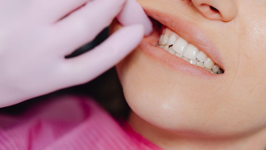
Lower wisdom teeth carry more nerve-related risk because the inferior alveolar nerve runs directly through the lower jawbone, often winding right between or beneath the roots of these teeth. Upper wisdom teeth sit far from any major nerve pathway, so the same concern rarely applies to them.
Lower wisdom tooth removal carries higher nerve injury risk than upper wisdom tooth removal because the mandibular canal, which houses the inferior alveolar nerve, frequently sits in direct contact with lower third molar roots.
If you’re planning a wisdom tooth extraction in Easton, PA and your dentist has flagged your lower wisdom tooth as “close to the nerve,” you’re not looking at a rare or unusual finding. It’s a documented anatomical pattern that shows up on X-rays and CBCT scans every day in oral surgery practices. Knowing why this happens, and what dentists do about it, makes the treatment plan easier to understand and puts the risk in proper context.
How Nerve Location Changes Lower Wisdom Tooth Removal
The lower jaw contains a bony canal that carries the inferior alveolar nerve along its length, and this canal sits close to where lower wisdom tooth roots develop. The upper jaw has no equivalent structure near the wisdom tooth site, which is why nerve injury conversations center almost entirely on the bottom teeth.
Which Nerves Lie Near Lower Wisdom Teeth?
Two nerves matter most in lower wisdom tooth surgery: the inferior alveolar nerve and the lingual nerve. The inferior alveolar nerve travels through the mandibular canal, a bony tunnel inside the lower jaw, supplying sensation to the lower lip, chin, and teeth on that side. The lingual nerve runs along the inner surface of the jawbone near the tongue, unprotected by bone in that stretch, which makes it vulnerable during soft tissue incisions.
In many patients, the roots of an impacted lower wisdom tooth sit within a millimeter or two of the mandibular canal, and in some cases the roots wrap around it or the canal notches into the root surface.
Why Upper Wisdom Teeth Have Different Anatomical Risks
Upper wisdom teeth face a different set of anatomical neighbors, and major nerves aren’t among them. The main structure of concern above is the maxillary sinus, which sits close to upper molar roots and can be affected during extraction, but sinus involvement doesn’t carry the same sensory nerve injury profile.
Upper jawbone is also less dense and more porous than the lower jaw, so upper wisdom teeth tend to extract with less surgical force and shorter procedure times.
What Nerve Injury Can Feel Like
Nerve injury after lower wisdom tooth extraction usually shows up as altered sensation rather than pain. Patients describe numbness, tingling, or a “fat lip” feeling in the lower lip, chin, or tongue on the affected side.
This condition, known clinically as paresthesia, resolves within weeks in most cases as nerve tissue recovers from stretching or bruising during surgery. Permanent nerve injury is uncommon, and dentists weigh this risk directly against the risks of leaving a problematic tooth in place.
Why Some Lower Wisdom Teeth Carry Greater Extraction Risk
Some lower wisdom teeth pose more nerve risk than others, and the difference comes down to how the tooth sits in the jaw and how it was imaged before treatment. Impaction angle, root shape, bone density, and the imaging method used all shift the risk profile for an individual patient.
How Impaction Angle and Root Shape Affect Risk
A tooth angled toward the nerve canal, or one with curved or splayed roots wrapping around it, raises extraction difficulty. Mesioangular and horizontal impactions, where the tooth leans forward or lies on its side, often place roots in closer contact with the mandibular canal than a vertically positioned tooth.
Root shape adds another layer. Fused, hooked, or unusually long roots are harder to remove cleanly and increase the chance of the surgeon needing to work near or around nerve tissue.
Why Dense Mandibular Bone Makes Surgery More Complex
Lower jawbone is denser and less flexible than upper jawbone, which limits how easily a tooth can be sectioned and removed. This density means more force, more drilling, and longer procedure times, all of which raise the technical demands of staying clear of the nerve canal.
Impaction rates are also higher in the lower jaw, so surgical extraction (rather than a simple extraction) is common, adding another variable to the risk equation.
How X-Rays and CBCT Scans Assess Nerve Proximity
A standard panoramic X-ray (OPG) is the first step in evaluating nerve proximity, and it can show warning signs like darkening of the root or interruption of the canal’s white outline. When an OPG raises concern, a CBCT scan provides a 3D view that shows whether the roots and canal are truly touching or just overlapping in a two-dimensional image.
CBCT imaging gives the surgeon precise information on canal position relative to the roots, which directly shapes the surgical approach chosen.
How Dentists Manage a Tooth Close to a Nerve
Dentists manage nerve-proximity cases by choosing between extraction, a modified surgical technique, or continued monitoring, based on imaging findings and symptoms. The decision balances the risk of nerve injury against the risk of leaving the tooth untreated.
When Monitoring May Be Safer Than Extraction
A wisdom tooth causing no pain, infection, or damage to neighboring teeth can sometimes be left in place and watched over time. This applies to fully impacted teeth with no signs of disease, where imaging shows close nerve contact and extraction risk outweighs the benefit of removal.
Regular check-ups and periodic imaging track whether the tooth’s position or condition changes.
Coronectomy and Other Risk-Reducing Options
A coronectomy removes the crown of the tooth while deliberately leaving the roots in place, avoiding disturbance to the nerve canal entirely. This technique suits cases where CBCT confirms the roots are in direct contact with the mandibular canal and full extraction would carry meaningful nerve injury risk.
Other risk-reducing approaches include sectioning the tooth into pieces for gentler removal and adjusting the surgical angle based on CBCT findings.
When Referral to an Oral Surgeon Is Appropriate
A general dentist refers to an oral surgeon when imaging shows a high-risk relationship between the tooth and the nerve canal. Oral surgeons have specialized training and equipment for managing close-contact cases, including microsurgical techniques and experience with coronectomy.
Referral is also common when a patient’s tooth position, root anatomy, or medical history adds complexity beyond routine extraction.
Careful Imaging and Planning Support Safer Treatment
Nerve proximity explains most of the added risk in lower wisdom tooth removal, and it stems from simple anatomy: the mandibular canal runs through the lower jaw near the roots, while the upper jaw has no comparable nerve pathway nearby. Impaction angle, root shape, and bone density all influence how close a call the extraction becomes.
Imaging drives every decision in these cases. A panoramic X-ray flags concern, and a CBCT scan clarifies whether the roots and nerve canal are truly in contact. That information shapes whether a dentist recommends extraction, coronectomy, or monitoring.
Nerve injury remains uncommon, and most cases of temporary numbness resolve within weeks. Discussing your imaging results directly with your dentist or oral surgeon gives you the clearest picture of your own risk and the safest path forward.